Watershed
The Watershed command separates objects that are close together or touching by treating pixel intensities as altitude and finding the valleys between peaks. It is placed after Connected Components.
When to use
Section titled “When to use”Use Watershed when high object density causes nearby objects to be detected as a single merged region after thresholding. Typical scenarios:
- Dense vesicle populations
- Touching cell nuclei
Parameters
Section titled “Parameters”| Parameter | Description |
|---|---|
| Maximum finder tolerance | Controls sensitivity to local intensity peaks. Range: 0.0–1.0. Lower values detect more (smaller) peaks and split more aggressively; higher values require more prominent peaks. |
How it works
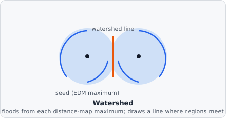
Section titled “How it works”- The Euclidean distance transform of the binary mask is computed - every foreground pixel’s value becomes its distance to the nearest background pixel, so the “deepest” interior of each blob becomes a local peak.
- Local maxima in the distance map are found; nearby maxima that don’t stick up above their connecting ridge by more than the tolerance are merged into one, which is what keeps noisy, near-flat peaks from over-splitting a single round object.
- Each surviving maximum is a seed; the mask floods outward from every seed simultaneously, growing downhill.
- Where two floods meet, a one-pixel-wide watershed line is drawn, splitting the original merged region into separate objects.
This is a faithful port of ImageJ’s Process > Binary > Watershed (its MaximumFinder class applied to the distance map), so results should match ImageJ pixel-for-pixel for the same input.
Background
Section titled “Background”The distance-map/immersion approach to watershed segmentation was formalized by Luc Vincent and Pierre Soille, “Watersheds in Digital Spaces: An Efficient Algorithm Based on Immersion Simulations,” IEEE Transactions on Pattern Analysis and Machine Intelligence, vol. 13, no. 6, pp. 583-598, 1991. The specific tolerance-based maximum-merging strategy ported here originates in ImageJ/Fiji - see Schindelin et al., “Fiji: An Open-Source Platform for Biological-Image Analysis,” Nature Methods, vol. 9, pp. 676-682, 2012.